
Introduction to Ultrasonography
Ultrasonography, also known as ultrasound imaging, is a medical diagnostic technique that uses high-frequency sound waves to produce images of structures within the body. It is a non-invasive, safe, and widely used method to visualize muscles, tendons, blood vessels, organs, and other internal structures.
Basic Principles of Ultrasonography
Sound Waves and Echoes
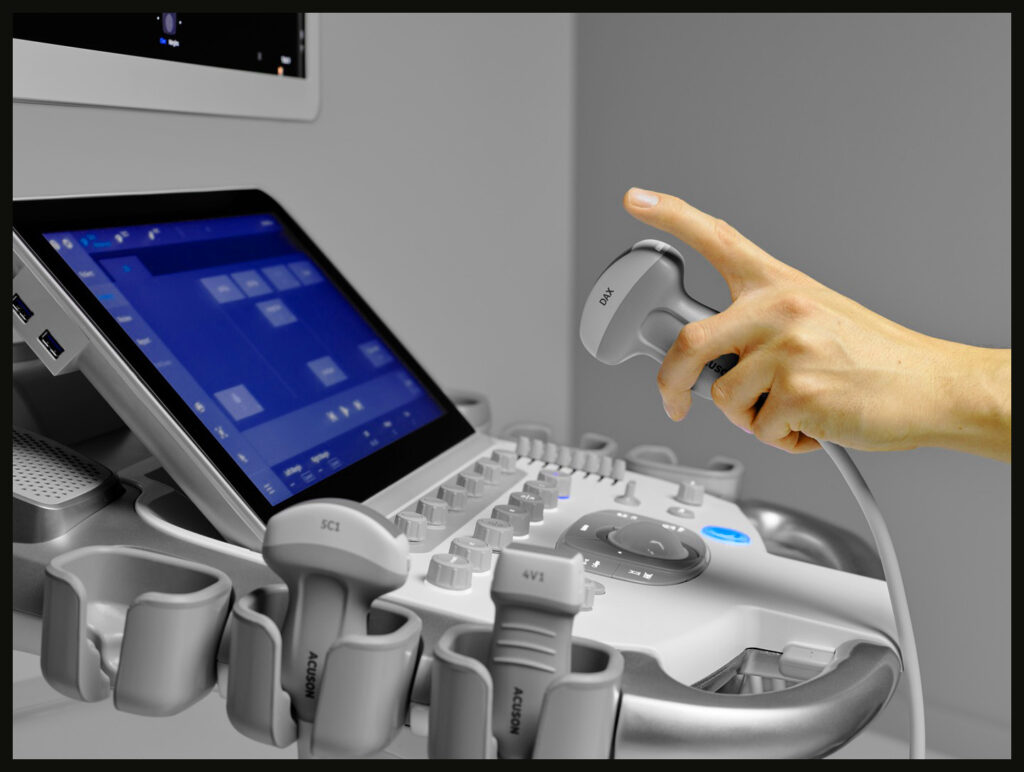
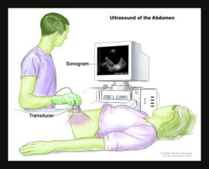
Ultrasonography relies on sound waves in the frequency range of 1 to 20 megahertz (MHz). A device called a transducer emits these sound waves, which travel into the body and reflect off internal structures. The returning echoes are detected by the transducer and converted into electrical signals. These signals are then processed to create visual images.
Image Formation
The images produced in ultrasonography are based on the varying densities of tissues and the way sound waves interact with them. Tissues of different densities reflect sound waves differently, allowing the creation of detailed images. The time it takes for the echoes to return to the transducer helps determine the depth and structure of the tissues being examined.
Types of Ultrasonography
2D Ultrasound
2D ultrasound is the most common type, producing flat, two-dimensional images of the internal structures. It is widely used in obstetrics, gynecology, cardiology, and general abdominal imaging.
3D Ultrasound
3D ultrasound technology builds upon 2D ultrasound by capturing multiple images from different angles and using computer software to create a three-dimensional image. This type of ultrasound provides more detailed and spatially accurate images, useful in obstetrics for viewing fetal development and in various other diagnostic applications.
4D Ultrasound
4D ultrasound, also known as dynamic 3D ultrasound, adds the dimension of time to 3D imaging. It creates real-time video images, allowing the observation of movement within the body, such as a fetus moving in the womb or blood flow through the heart.
Doppler Ultrasound
Doppler ultrasound measures and visualizes blood flow within vessels using the Doppler effect, which is the change in frequency of sound waves as they reflect off moving objects. There are different types of Doppler ultrasound:
- Color Doppler: Uses color to represent the direction and velocity of blood flow.
- Power Doppler: Provides more sensitive detection of blood flow, especially in small vessels, but does not show direction.
- Spectral Doppler: Displays blood flow measurements graphically, showing changes in velocity over time.
Applications of Ultrasonography
Obstetrics and Gynecology
Ultrasonography is extensively used in obstetrics for monitoring fetal development, assessing fetal health, and diagnosing potential issues during pregnancy. Key applications include:
- Early Pregnancy Assessment: Confirming pregnancy, determining gestational age, and assessing for multiple pregnancies.
- Anatomical Survey: Evaluating fetal anatomy for abnormalities.
- Growth Monitoring: Measuring fetal growth and estimating weight.
- Placental Assessment: Checking the position and health of the placenta.
- Amniotic Fluid Assessment: Measuring the amount of amniotic fluid.
Cardiology
Cardiac ultrasonography, or echocardiography, is vital for assessing heart function and diagnosing cardiovascular diseases. Key uses include:
- Ejection Fraction Measurement: Evaluating the percentage of blood the heart pumps out with each contraction.
- Valve Assessment: Checking for valve abnormalities, such as stenosis or regurgitation.
- Structural Analysis: Examining heart chamber sizes, wall thickness, and detecting congenital heart defects.
- Blood Flow Evaluation: Using Doppler ultrasound to measure blood flow and detect abnormalities.
Abdominal Imaging
Ultrasound is used to visualize abdominal organs, including the liver, gallbladder, kidneys, pancreas, and spleen. It helps diagnose conditions like:
- Gallstones: Detecting stones in the gallbladder or bile ducts.
- Liver Disease: Assessing liver size, structure, and detecting tumors or cysts.
- Kidney Stones: Identifying stones and other kidney abnormalities.
- Pancreatic Issues: Evaluating the pancreas for signs of inflammation, tumors, or cysts.
Musculoskeletal Imaging
Ultrasound is increasingly used in musculoskeletal imaging to assess muscles, tendons, ligaments, and joints. Applications include:
- Tendon and Muscle Injuries: Diagnosing tears, strains, and other soft tissue injuries.
- Joint Inflammation: Detecting inflammation and fluid accumulation in joints.
- Nerve Entrapments: Identifying conditions like carpal tunnel syndrome.
- Guided Injections: Assisting in the accurate placement of needles for injections or aspirations.
Vascular Imaging
Vascular ultrasound is used to examine blood vessels and diagnose vascular conditions. Key applications include:
- Deep Vein Thrombosis (DVT): Detecting blood clots in deep veins, typically in the legs.
- Carotid Artery Disease: Assessing the carotid arteries for blockages or narrowing.
- Aneurysms: Identifying abnormal bulging in blood vessel walls.
- Peripheral Artery Disease (PAD): Evaluating blood flow in peripheral arteries to detect blockages or narrowing.
Advantages of Ultrasonography
Non-Invasive and Safe
Ultrasonography is non-invasive, meaning it does not require incisions or injections. It is also considered safe, as it does not use ionizing radiation like X-rays or CT scans. This makes it suitable for a wide range of patients, including pregnant women and children.
Real-Time Imaging
Ultrasound provides real-time imaging, allowing for dynamic assessment of structures and functions within the body. This is particularly useful in procedures that require immediate feedback, such as guiding needle placements or monitoring fetal movements.
Portability and Accessibility
Ultrasound machines are relatively portable compared to other imaging modalities, such as MRI or CT scanners. This portability makes ultrasound accessible in various settings, including hospitals, clinics, and even remote locations. Handheld ultrasound devices further enhance accessibility.
Cost-Effective
Ultrasound is generally more cost-effective than other imaging techniques. It is often used as a first-line diagnostic tool due to its affordability and the ability to provide quick and accurate assessments.
Limitations of Ultrasonography
Operator Dependence
The quality and accuracy of ultrasound imaging can be highly dependent on the skill and experience of the operator. Proper training and expertise are crucial to obtain reliable and diagnostically useful images.
Limited Penetration and Resolution
Ultrasound waves may have limited penetration through dense tissues or air-filled structures, such as bone and lungs. This can result in reduced image quality and diagnostic challenges. Additionally, the resolution of ultrasound images may be lower than that of other imaging modalities like MRI or CT.
Artifacts
Ultrasonography can produce artifacts—false or misleading visual elements—that can complicate image interpretation. Common artifacts include shadowing, enhancement, and reverberation. Understanding and recognizing these artifacts are essential for accurate diagnosis.
Technological Advances in Ultrasonography
Contrast-Enhanced Ultrasound (CEUS)
CEUS involves the use of contrast agents to improve the visualization of blood flow and tissue vascularity. It enhances the ability to detect and characterize lesions, especially in the liver, kidneys, and other organs.
Elastography
Elastography measures tissue stiffness and elasticity, providing additional diagnostic information. It is particularly useful in evaluating liver fibrosis, thyroid nodules, and breast lesions. There are two main types:
- Strain Elastography: Measures tissue deformation in response to manual compression.
- Shear Wave Elastography: Uses acoustic radiation force to generate shear waves and measure tissue stiffness quantitatively.
Artificial Intelligence (AI) and Machine Learning
AI and machine learning are being integrated into ultrasound imaging to enhance image acquisition, interpretation, and diagnosis. These technologies can assist in identifying abnormalities, improving diagnostic accuracy, and reducing operator dependence.
Portable and Handheld Devices
Advances in technology have led to the development of portable and handheld ultrasound devices. These devices are smaller, more affordable, and offer improved accessibility, particularly in remote or resource-limited settings. They are useful for point-of-care diagnostics, emergency medicine, and telemedicine applications.
Conclusion
Ultrasonography is a versatile, safe, and essential imaging modality in modern medicine. Its ability to provide real-time, non-invasive, and cost-effective imaging makes it invaluable in various clinical settings. While it has some limitations, ongoing technological advancements continue to enhance its capabilities and expand its applications. As ultrasonography technology evolves, it will remain a cornerstone of diagnostic imaging, offering critical insights into the human body and aiding in the diagnosis and management of numerous medical conditions.